Cardiac Output Monitoring: A Contemporary Assessment and Review
Authors, Journal, Affiliations, Type, DOI
- Authors: Robert H. Thiele, MD; Karsten Bartels, MD; Tong J. Gan, MD, MHS, FRCA
- Journal: Critical Care Medicine 2015;43(1):177–85 (January 2015). Copyright © 2014 Society of Critical Care Medicine and Lippincott Williams & Wilkins
- Affiliations: Department of Anesthesiology, University of Virginia, Charlottesville; Department of Anesthesiology, University of Colorado Denver, Aurora; Department of Anesthesiology, Stony Brook University, Stony Brook, NY
- Type: Narrative/systematic review of cardiac output monitoring technology, organised by underlying physical principle. PubMed search yielding 1,526 sources, with systematic extraction of animal model, number of independent measurements, and correlation between techniques for each study. Appendix 1 (Supplemental Digital Content) tabulates all reviewed data
- DOI: 10.1097/CCM.0000000000000608
- Disclosures: Dr. Thiele's institution consulted for Teleflex (Airway Advisory); Dr. Gan consulted for Merck, Edwards, Covidien and Deltex, and his institution received grant support from Fresenius; Dr. Bartels reported no conflicts. Supported in part by departmental sources
Overview
A principle-by-principle review of every commercially relevant way to measure cardiac output, written to let clinicians choose a device on the basis of what it actually measures rather than what it claims. Its central verdict: thermodilution is the most accurate technique against experimental reference standards; Doppler-based methods approach it in accuracy while being less invasive and offering beat-to-beat trending, at the cost of dependence on beam alignment and knowledge of aortic cross-sectional area; bioimpedance is less accurate; and the newer classes — pulse contour, partial rebreathing, pulse wave velocity — rest on far fewer studies, are mostly validated against thermodilution rather than a true reference standard, and in the majority of studies show decreased accuracy. The review is valuable for its equations: it derives each device family from its physical basis, and those derivations are what distinguish the techniques and explain their failure modes. All ten numbered equations are transcribed in full below, together with their original symbol definitions.
Keywords
Cardiac output; echocardiography; goal-directed therapy; hemodynamic monitoring; minimally invasive monitoring; pulmonary artery catheter
The Ten Equations of Cardiac Output Measurement
Every numbered equation in the review, transcribed and reproduced. The source PDF renders these as typeset images rather than selectable text, so each is given both as transcribed text (searchable) and as the original image (visually faithful).
Fick principle — Equation 1
Described by Adolf Eugen Fick (1829–1901) as the relationship between cardiac output, oxygen uptake, and the arteriovenous oxygen content difference. This is the foundation of the oximetric Fick reference method.
CO = V̇o₂ / (Cao₂ − Cvo₂)

- Symbols: CO = cardiac output; V̇o₂ = oxygen uptake; Cao₂ − Cvo₂ = difference between arterial and venous blood oxygen content
Indicator dilution — Equation 2 (Stewart–Hamilton)
Stewart's "indicator-dilution" method relies on injecting an indicator (e.g. dye) and measuring a time–concentration curve. Flow is calculated from derivatives of the Conservation of Indicator Principle, known as the Stewart–Hamilton equations.
m = Q ∫₀^∞ c(t)dt

- Symbols: m = mass of indicator injected; Q = flow; c(t) = function describing change in concentration over time
Thermodilution — Equation 3 (Swan–Ganz)
Fegler adapted Stewart's technique using temperature as the indicator. To apply this in Swan thermodilution the additional factors below must be considered.
CO = V_I(T_B − T_I) K₁ × K₂ / ∫₀^∞ ΔT_B(t)dt

- Symbols: V_I = injectate volume; T_B = blood temperature; T_I = injectate temperature; K₁ = density factor (specific heat × specific gravity of injectate, divided by the product of specific heat and gravity of blood); K₂ = computation constant accounting for catheter dead space, heat exchange in transit, and injection rate; the product is divided by the change in blood temperature over time, ∫₀^∞ ΔT_B(t)dt
- The three assumptions this technique makes: (i) measuring the temperature curve over a finite rather than indefinite period does not meaningfully affect the estimate of its integral; (ii) injectate and bloodstream are perfectly mixed; (iii) the measured temperature difference is accurate. These fail in tricuspid regurgitation, frequent repeated measurements, low-flow states, and rapid temperature change after cardiopulmonary bypass
Doppler ultrasound — Equation 4
Relates the frequency change of a returning ultrasound beam to the velocity of a moving reflector, via the speed of sound in tissue and the angle of incidence.
v = cΔf / (2f₀ cos(θ))

- Symbols: v = velocity of the moving reflector; c = speed of sound in tissue; Δf = frequency change of the returning beam; f₀ = frequency of the incident beam; θ = angle of incidence
- To obtain stroke volume the velocity of blood flow must be integrated over one entire heartbeat (the "velocity time integral") and multiplied by cross-sectional area. Two neglected error sources: cross-sectional area changes over the cardiac cycle, and the measured velocity is assumed identical at all points in the vessel (a "flat" velocity profile)
CO₂-based Fick — Equation 5
Carbon dioxide can measure CO using a modified version of the Fick equation.
CO = V̇co₂ / (Cpaco₂ − Csaco₂)

- Symbols: CO = cardiac output; V̇co₂ = production of carbon dioxide; Cpaco₂ = concentration of CO₂ in the pulmonary arterial compartment; Csaco₂ = concentration of CO₂ in the systemic arterial compartment
Partial rebreathing — Equations 6, 7, 8
The partial rebreathing technique assumes CO does not change between periods of non-rebreathing (minimal dead space) and partial rebreathing (dead space added to the ventilator circuit). CO in both states can be written as two equations:
CO = V̇co₂,non / (Cpaco₂,non − Csaco₂,non)
   = V̇co₂,rebr / (Cpaco₂,rebr − Csaco₂,rebr)
(6)

Combining the two states gives:
CO = (V̇co₂,non − V̇co₂,rebr) / [ (Cpaco₂,non − Csaco₂,non) − (Cpaco₂,rebr − Csaco₂,rebr) ]
(7)
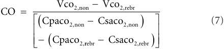
If one assumes Cpaco₂ does not change between the non-rebreathing and partial rebreathing periods, the equation simplifies:
CO = V̇co₂,non − V̇co₂,rebr / (Csaco₂,rebr − Csaco₂,non)
(8)

- Fidelity note on equation 8: as typeset in the paper the numerator
V̇co₂,non − V̇co₂,rebrcarries no parentheses, so read literally the expression parses asV̇co₂,non − (V̇co₂,rebr / (…)). The intended meaning is a single quotient with the grouped difference as numerator, and the image above preserves the original typesetting so the discrepancy is visible. Flagged rather than silently corrected - Csaco₂ estimation: derived from haemoglobin concentration and arterial CO₂ partial pressure. Nunn iso-shunt plots (relating Q_s/Q_T to Pao₂ and Fio₂) estimate Q_s/Q_T, accounting for intrapulmonary shunts
Pulse contour analysis — Equation 9 (PiCCO stroke volume)
Frank's Windkessel model of blood flow underlies most early attempts to derive CO from the arterial pulse contour; the aorta acts as a capacitor and the systemic arterioles as a resistor (the "two-element Windkessel model"). The PiCCO system treats compliance as a dynamic variable dependent on pressure.
SV = k ∫_end-diastole^end-systole [ P(t)/SVR + C(p) × dP/dt ] dt
(9)

- Symbols: SV = stroke volume; k = calibration constant requiring transpulmonary thermodilution to determine; P(t) = arterial pressure waveform; SVR = systemic vascular resistance; C(p) = pressure-dependent compliance, based on waveform analysis distal to the dicrotic notch; dP/dt = instantaneous pressure change
- The PiCCO modifications aim to account better for the fraction of ventricular output stored in capacitance vessels
Pulse wave velocity — Equation 10 (Moens–Korteweg)
Describes the velocity of a pulse wave through an elastic tube using the tube's elasticity, wall thickness and diameter, and the density of the fluid within it.
V = k(Eh / ρD)^(1/2)
(10)

- Symbols: V = pulse wave velocity; E = tube elasticity (vessel wall elastic modulus); h = wall thickness; ρ = density of the fluid within the tube; D = tube diameter; k = constant
- The technique's two assumptions: stroke volume is proportionate to pulse pressure; and pulse pressure is related to pulse wave velocity (or pulse wave transit time, PWTT)
Key Takeaways
Methods
- A systematic PubMed search identified all published articles describing the use of CO monitors. For historical consistency the authors included studies using regression analysis, while not neglecting those using bias and limits-of-agreement analysis
- Animal studies were included where available: although less persuasive than human data in one sense, they add value because investigators can achieve a wider range of haemodynamic conditions — particularly useful given the range-dependence of the regression approach
- The methodological problem the review is built on: criticisms of linear regression include its dependence on a wide range of measurements and the inordinate effect of outliers. When measured values are relatively stable (as in humans), methods that agree closely can exhibit very little correlation
Thermodilution
- The PAC era began when thermodilution was combined with the balloon-tipped pulmonary artery catheter by Swan and Ganz, making CO measurement a clinical reality by the early 1970s. A succession of large prospective RCTs then failed to show mortality improvement in critically ill patients, leading to declining PAC use
- Thermodilution has been validated in a greater number and variety of experimental models than any other clinically available technique, including ex vivo mechanical models and reference standards in animals and humans. Its consistent accuracy plus its first-to-market position explain why thermodilution PACs became the clinical reference standard for later comparison studies. The precision error of PACs is 13% for triplicate readings
- Continuous thermodilution catheters use a heating element rather than room-temperature saline and appear comparably accurate, with a time lag of up to 5 minutes
- The PAC is unique in also measuring pulmonary artery and "occlusion" pressure — though the ability of practising physicians to interpret PA tracings is in doubt
- Swan thermodilution measures between the right atrium and pulmonary artery, so it represents only right-sided CO and dismisses shunt
Ultrasound-based techniques
- Doppler-based devices have been validated in ex vivo flow models and against reference standards in animals and humans, suggesting a slight decrease in accuracy versus thermodilution — but "the magnitude of this difference is of questionable clinical significance"
- Major advantage: noninvasive, with beat-to-beat measurement and excellent trending
- Esophageal Doppler images the descending thoracic aorta. Unlike the suprasternal approach it cannot directly measure CO, since ~30% of CO is diverted proximal to the descending thoracic aorta. Comparisons with Fick methods show correlations just slightly worse than expected from a PAC
- Modern esophageal Doppler devices also provide "corrected flow time" and stroke volume variation (SVV) as fluid-responsiveness endpoints. Esophageal Doppler cannot be more accurate than thermodilution, because its stroke-distance-to-stroke-volume algorithm is derived from a Doppler/thermodilution database
Bioimpedance and bioreactance
- Thoracic electrical bioimpedance (TEB) assumes thoracic electrical resistance relates to intrathoracic blood volume, and that impedance changes are exclusively a function of blood volume change. It is highly sensitive to electrode positioning, pulmonary oedema and electrical noise. Results suggest bioimpedance devices are less accurate than thermodilution or Doppler; the propensity for interference in the operating room suggests alternatives should be considered when available
- Electrical velocimetry assumes thoracic conductivity relates to the velocity of aortic blood flow rather than blood volume. Available data are insufficient to recommend widespread use
- Bioreactance was developed to reduce errors from electrode positioning, body size, temperature and humidity; intrathoracic blood behaves as both capacitor and inductor, and these properties affect the phase shift between applied and received voltage. Compared with thermodilution, the mean weighted correlation coefficient was 0.80 across three studies containing 171 comparisons. The only available device is the NICOM (Cheetah Medical), which also estimates SVV
Partial rebreathing
- The technique has primarily been validated against thermodilution in animals, with one transit-time flow probe study. Human comparisons include more than 800 paired measurements from over 250 patients in at least nine studies
- Accuracy appears related to tidal volume and shunt fraction, and the technique has a relatively long response time
- The NICO system (Philips Respironics) is the only commercially available system using partial rebreathing. To be completely noninvasive it assumes the difference between end-tidal CO₂ and Csaco₂ is approximately 6 mm Hg
Pulse contour analysis
- Two classes exist: calibrated devices (recalibrating periodically from a second technique, e.g. transpulmonary thermodilution or lithium dilution) and uncalibrated devices
- Calibrated: transpulmonary thermodilution (PiCCO, Pulsion Medical Systems) and lithium dilution (LiDCO Plus / PulseCO, LiDCO Group), the latter using characteristic impedance with a transfer function to estimate aortic pressures. Lithium injected centrally is a reliable indicator; peripheral injection reduces accuracy
- Uncalibrated: the empiric approach (FloTrac, Edwards Lifesciences) derives a mathematical rather than physical model and "likely comes at the expense of decreased accuracy"; and PRAM (pressure recording analytical method), based on volumetric changes occurring primarily in the radial direction, studied by a single group so requiring further validation
- Calibrated vs uncalibrated: a simultaneous comparison of LiDCO, PiCCO and FloTrac against intermittent thermodilution showed narrower limits of agreement for the calibrated devices. Accuracy of all three suffers during haemodynamic instability; the majority of data suggest calibrated devices outperform uncalibrated
- Pulse contour devices as a class must be used cautiously in aortic insufficiency, which alters arterial waveform shape
Photoplethysmographic
- The PPG waveform relates to arterial pressure and may relate to stroke volume in idealized conditions, but this relationship is not clinically useful in acute care
- Peñáz's "volume clamp" technique monitors finger arterial blood volume with an infrared transmitter/receiver connected to an inflatable finger bladder driven by feedback. Maintaining the artery "unstretched" allows continuous finger pressure measurement. Agreement with invasive blood pressure is reasonable, but the Finapres and Nexfin stroke-volume estimates cannot yet be recommended for reliable CO measurement
Pulse wave velocity
- The esCCO (Nihon Kohden) estimates CO by incorporating PWTT (from ECG and pulse oximeter waveforms) and mean arterial pressure into stroke volume estimation, with proprietary details. Not FDA approved, marketed in Japan; has not demonstrated sufficient accuracy to recommend widespread clinical use
Conclusions
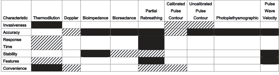
Figure 1 — the review's qualitative comparison of commercially available cardiac output monitoring technology, across calibrated and uncalibrated pulse contour, Doppler, bioimpedance, rebreathing and other classes. White = above average, lined = average, black = below average (or insufficient data to assess).
- Thermodilution is relatively accurate against reference standards such as the oximetric Fick method or electromagnetic and transit-time flow meters
- Doppler-based techniques have similar accuracy to thermodilution PACs and offer beat-to-beat monitoring with less invasiveness. However, direct comparisons between Doppler and thermodilution against experimental reference standards suggest thermodilution is more accurate
- The assumptions underlying Doppler- and thermodilution-based derivation should be known and considered in a given clinical context
- Bioimpedance devices do not correlate with direct Fick methods as closely as either thermodilution or Doppler. Electrical velocimetry and bioreactance need more data
- The literature for newer devices is significantly smaller and primarily based on comparison with thermodilution rather than a reference standard. Uncalibrated pulse contour devices appear to track CO changes when afterload is stable; calibrated devices are more accurate than uncalibrated in haemodynamically unstable patients but need periodic operator intervention. Partial rebreathing and pulse wave velocity devices have not demonstrated sufficient accuracy to justify widespread clinical use
Limitations of the document
Stated and evident in the review itself
- The review is built on a methodology its own authors criticise. The included studies predominantly used regression/correlation analysis, which the paper notes is range-dependent and outlier-sensitive; in humans, where CO is relatively stable, "methods which agree closely can exhibit very little correlation." The authors include these studies "for purposes of historical consistency" rather than because the method is sound
- No formal quality appraisal, no meta-analysis, no GRADE. This is a narrative synthesis organised by principle. Correlation coefficients are reported across studies without pooling, and heterogeneity in models, measurement counts and reference standards is not formally assessed
- The synthesis is qualitative. There is no quantitative estimate of accuracy for any device class; the comparative statements rest on counted directions of effect rather than pooled estimates
- Two of three authors have device-industry relationships (Teleflex; Merck, Edwards, Covidien, Deltex; institutional support from Fresenius), disclosed in the article. The devices discussed include products from those companies, and the review's conclusions are favourable to the more established — and more invasively monitored — technologies
- Publication date. 2015. The device landscape has moved substantially since; the newest classes described here (esCCO, and several pulse contour devices) are presented as immature, and no device released after 2015 is covered
Key Concepts Mentioned
- concepts/Cardiac-Output-Measurement — the technique-by-technique page this review supplies the equations for
- concepts/Right-Heart-Catheterization — the pulmonary artery catheter as both clinical and de facto experimental reference standard
- concepts/Pulmonary-Artery-Pulsatility-Index — another PAC-derived haemodynamic index
- concepts/Cardiopulmonary-Exercise-Testing — also estimates cardiac output, by the Fick principle, during exercise
- concepts/Intracardiac-Shunts — shunt is the specific error the Fick and thermodilution methods cannot see, and the reason Nunn iso-shunt plots are needed for Csaco₂ estimation
- Fick principle — the oximetric reference standard, and the basis for CO₂ rebreathing
- Stewart–Hamilton equation — the indicator-dilution foundation, including thermodilution
- Windkessel model — Frank's two-element model underlying pulse contour analysis
- Moens–Korteweg equation — pulse wave velocity through an elastic tube
- Velocity time integral — Doppler-derived stroke volume requires VTI × cross-sectional area
- Nunn iso-shunt plots — estimate intrapulmonary shunt fraction for Csaco₂
Key Entities Mentioned
- concepts/Cardiogenic-Shock — the clinical state in which CO measurement most often drives decisions
- concepts/Cardiogenic-Shock — contemporary shock staging alongside which CO monitors sit
- Adolf Eugen Fick (1829–1901) — described the relationship underlying the reference method
- Swan and Ganz — balloon-tipped pulmonary artery catheter and its thermodilution adaptation
- Stewart and Hamilton — indicator-dilution equations
- Fegler — adapted indicator dilution to temperature as the indicator
- Peñáz — "volume clamp" technique for continuous finger arterial pressure
- Frank — Windkessel model of blood flow
- Moens and Korteweg — pulse wave velocity equation
- Device manufacturers named: Pulsion (PiCCO), LiDCO Group (LiDCO Plus/PulseCO), Edwards Lifesciences (FloTrac), Vytech Health (PRAM), Cheetah Medical (NICOM), Philips Respironics (NICO), Nihon Kohden (esCCO), FMS (Modelflow), Deltex (esophageal Doppler)
Wiki Pages Updated
- Created: wiki/sources/co-ccmj-2015.md
- Updated: wiki/concepts/Cardiac-Output-Measurement.md
- Updated: wiki/sourceindex.md
- Updated: wiki/wikiindex.md